Principal Spectroscopy Equipment
Confocal Raman Microspectrometer (632.8 nm excitation)
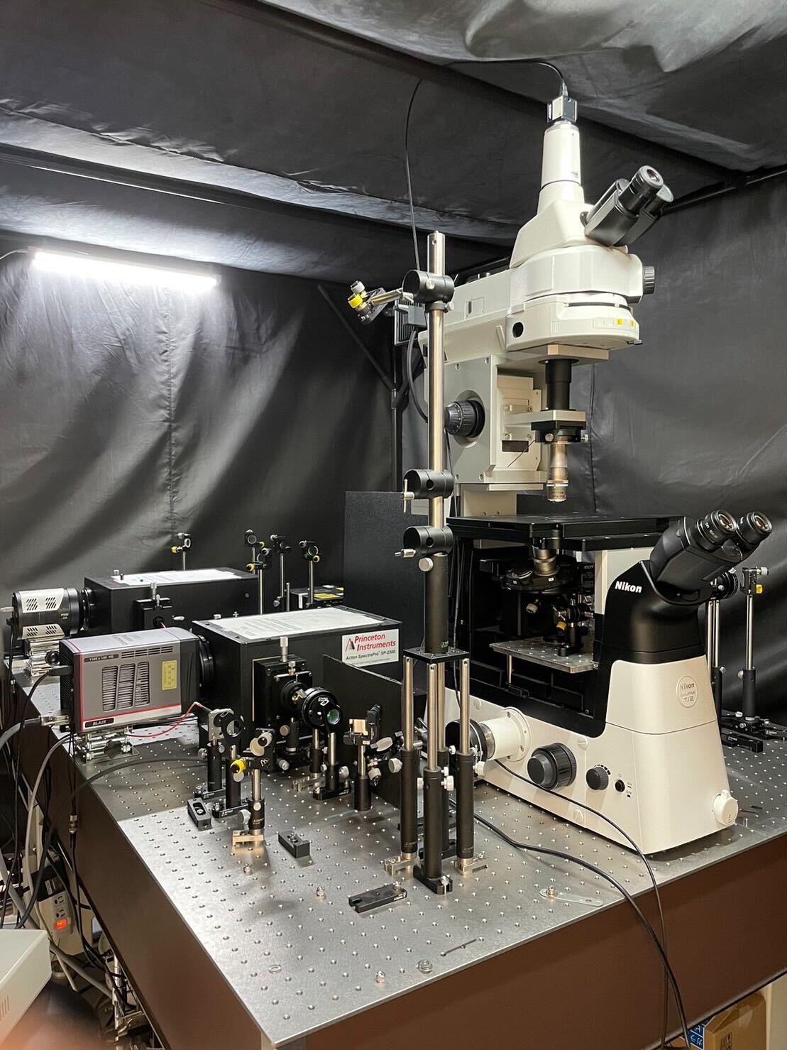
This laboratory-built instrument can measure Raman spectra of the sample such as a living cell under an inverted microscope (Nikon Ti2-U) with a submicrometer resolution. It uses the 632.8 nm output of a He–Ne laser as the Raman excitation light. The detector used is a TE-cooled CCD detector (Andor iDus DU401-BV) or an electron-multiplying CCD detector (Andor Newton DU970P-BVF). By incorporating volume Bragg grating notch filters that have a very narrow bandwidth, we are able to observe an extremely low-frequency region (down to ±5 cm-1) of Raman spectra. A 3-axis piezoelectric stage (Mad City Labs, Nano-LP100) can be mounted on the microscope stage for Raman imaging experiments.
Confocal Raman Microspectrometer (532 nm and Multi-Wavelength Excitation)
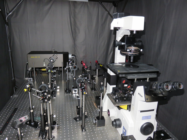
This Raman microscope is also laboratory-built. It is based on an Olympus inverted microscope (IX73). The excitation laser is an all-solid, optically pumped semiconductor laser (Coherent, Genesis CX532-2000SLM-CDRH) or wavelength tunable nanosecond OPO laser (EKSPLA, NT242), and the detector is an electrically cooled CCD detector (Andor, iDus DU401-BV). Like the 632.8 nm excited Raman microscope, this apparatus is also capable of measuring Raman spectra as low as ±5 cm-1. Using a precise temperature control unit for microscopy (Interference, VAHEAT) or a microscope cooling/heating stage (Linkam, 10021), we can measure Raman spectra at different temperatures.
Multimodal Nonlinear Microspectrometer
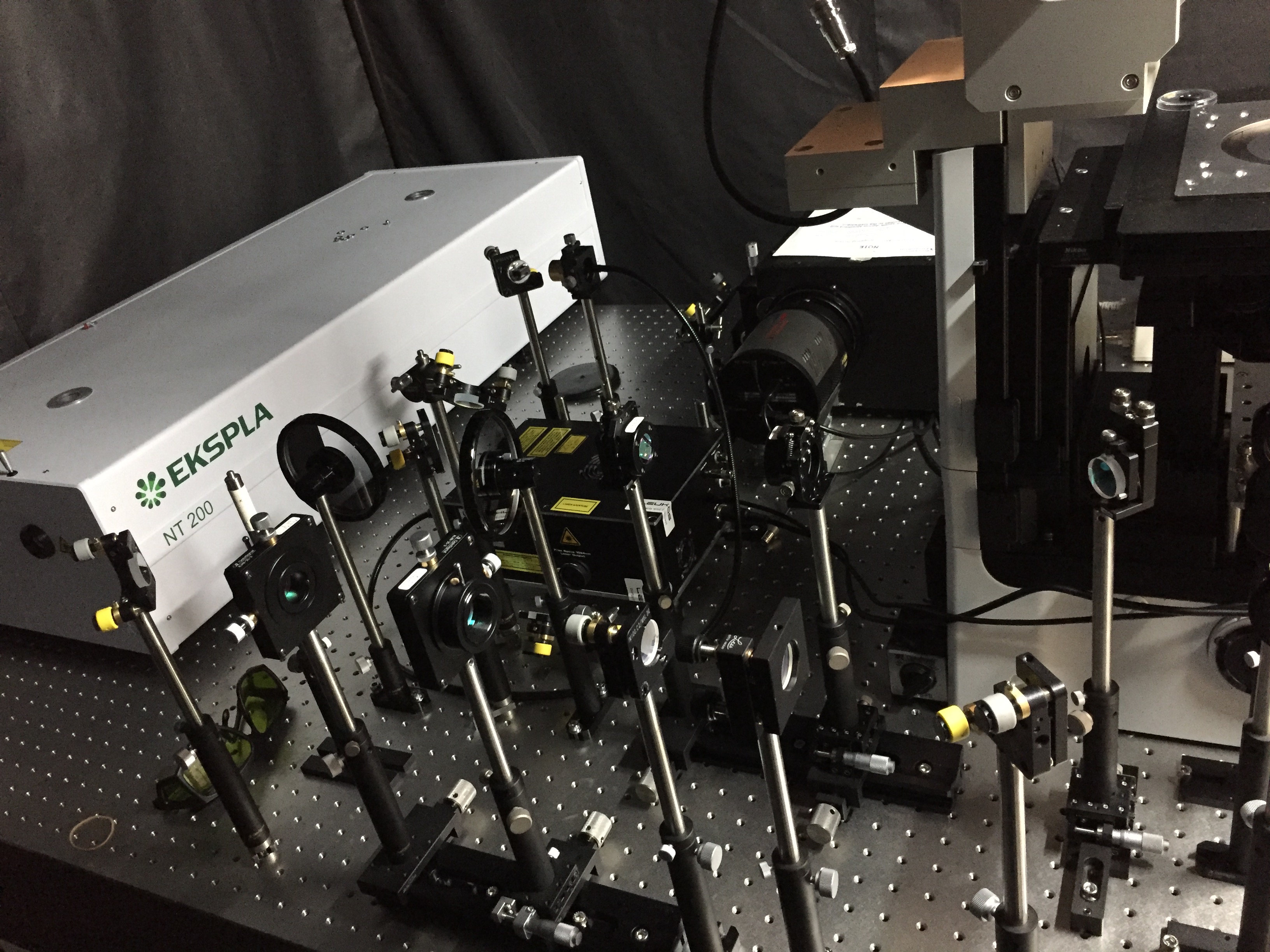
This microspectrometer utilizes coherent anti-Stokes Raman scattering (CARS), a third-order nonlinear optical process. Whereas the above two instruments use linear (spontaneous) Raman scattering, CARS is superior in sensitivity because it can generate coherent signal light with high efficiency, and therefore it can achieve faster imaging speed (~1 ms). Another important feature of this setup is the multiplex capability, in which many vibrational modes can be detected simultaneously using supercontinuum light in the near-infrared region generated from a photonic crystal fiber. In addition to CARS, this microspectrometer can also observe nonlinear optical signals such as second harmonic generation (SHG) and third harmonic generation (THG) that appear in the visible region.
Nano Raman Imaging Apparatus
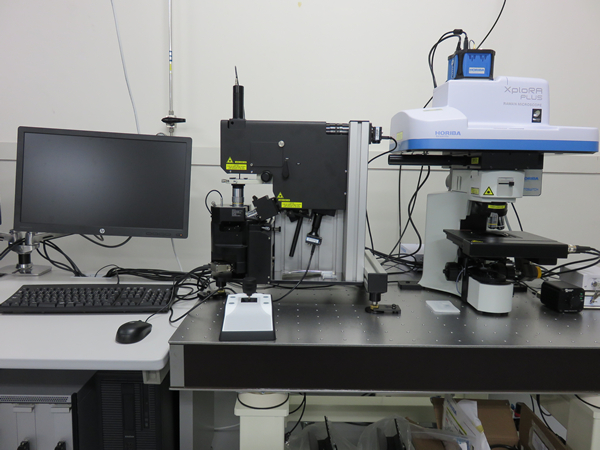
This state-of-the-art apparatus that combines an atomic force microscope (AFM) and an upright Raman microscope allows not only for Raman measurements synchronized with AFM topographic imaging but also for nanoscale chemical analysis by tip-enhanced Raman scattering (TERS) measurements. 532 nm and 638 nm lasers are implemented for Raman excitation. We use this apparatus to study membrane vesicles produced by bacterial cells as well as thin films of organic-inorganic hybrid perovskites and amyloid fibrils.
Infrared Electroabsorption Spectrometer
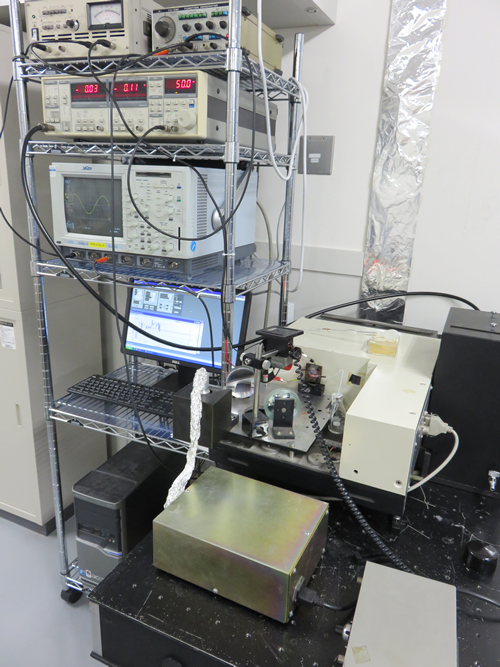
This spectrometer capable of detecting IR absorbance changes of ΔA ≈ 10-8 induced by application of an external electric field is unique to our laboratory. The high sensitivity is achieved by using a dispersive monochromator in combination with AC-coupled amplification, rather than a Fourier transform spectrometer. The detector used is a HgCdTe or an InSb infrared detector. With this apparatus, we are currently investigating the electric field effects of water in reverse micelles and of CH3NH3PbI3 thin films.
Other Equipment
FTIR Spectrometer (right), UV/VIS Spectrometer (left)
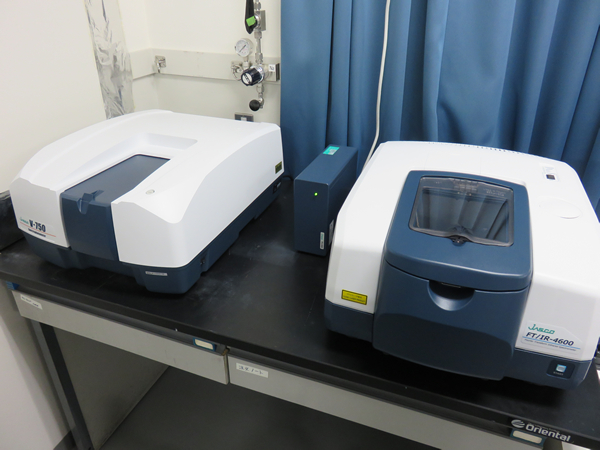
These are routinely used for steady-state measurements. An option of attenuated total reflection (ATR) with a ZnSe prism is available for the FTIR spectrometer.
Fluorescence Microscope
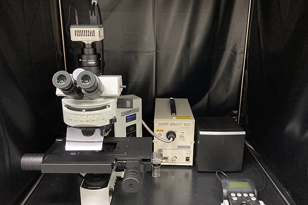
This is a microscope for fluorescence observation of cells and tissues with UV, B, and G excitation.
Nanoparticle Analyzer
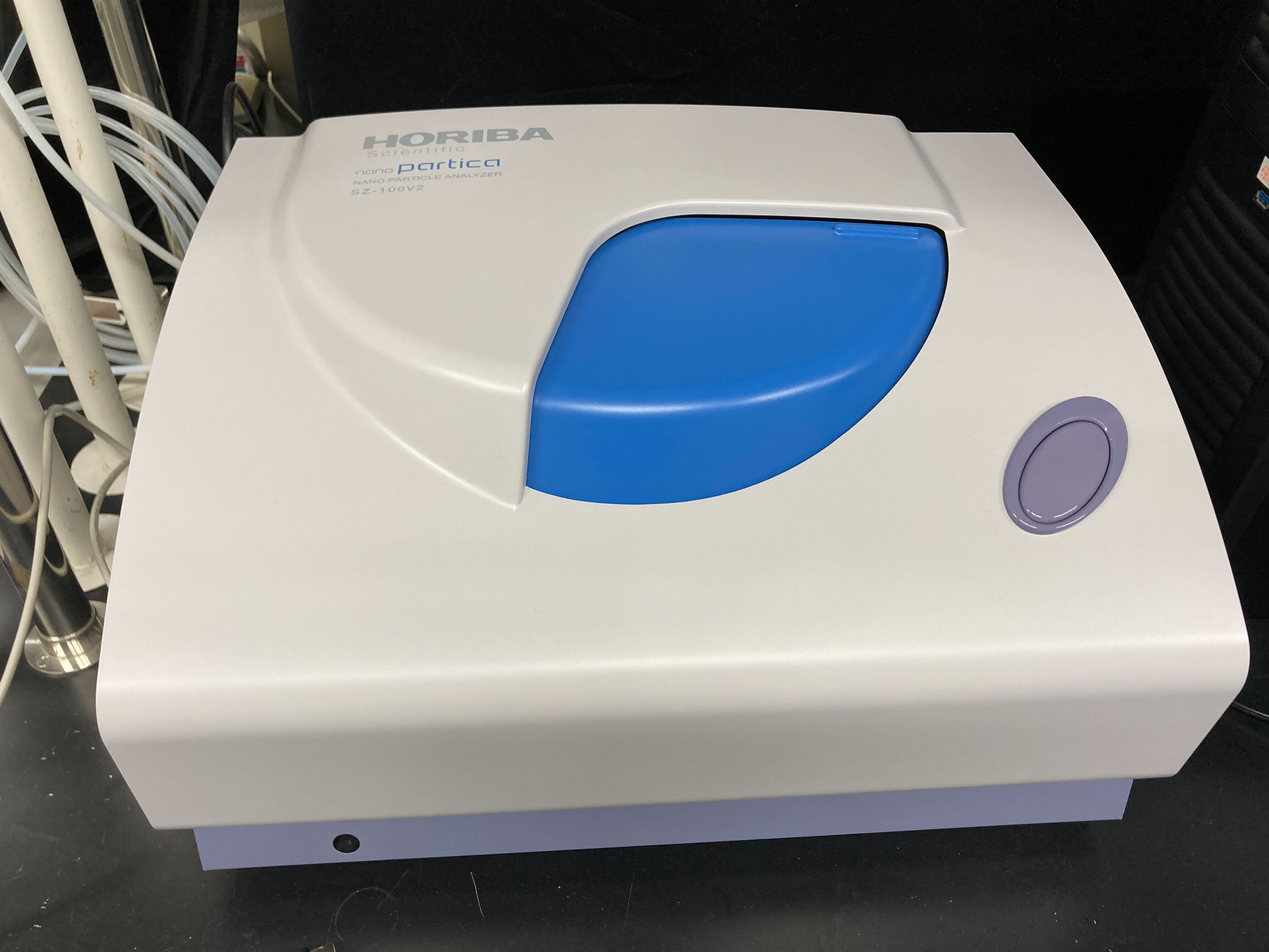
This equipment (HORIBA, SZ-100) measures the size distribution of nanoparticles such as metal nanoparticles and membrane vesicles based on dynamic light scattering measurements.
Cell Culture Facilities
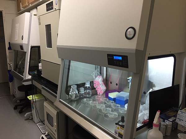
We have two clean benches, six shaking incubators with different operating temperature ranges, and two -80°C deep freezers. We also have an autoclave for sterilization, centrifuges, and microscopes for microbial observation. We culture various bacteria and fungi such as filamentous fungi and microalgae provided by our collaborators (RIKEN JCM, Nomura group at University of Tsukuba, Sakudani group at Tokushima University, Nojiri group at the University of Tokyo, Kawamukai group at Shimane University, etc.) and measure the cultured cells with the above-mentioned Raman microspectrometers and fluorescence microscope. All of these microorganisms are nonpathogenic, including environmental commensal bacteria.
Centrifuges
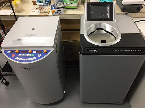
These instruments are used to separate and concentrate microbial cells and to concentrate and purify membrane vesicles produced by microbes. One of them (himac, CS120FNS) is an ultracentrifuge that can achieve a maximum rotation speed of 120,000 rpm and a maximum centrifugal acceleration of 771,000×g.
Ultrapure Water System
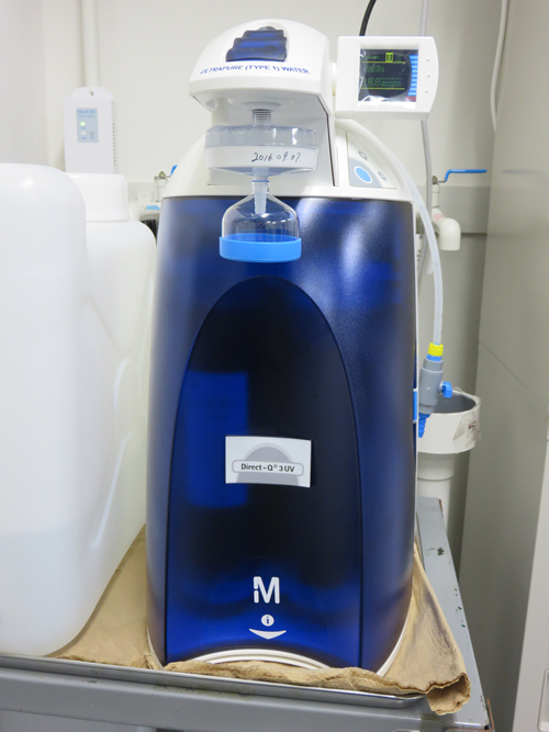
This equipment (Merck Millipore Direct-Q UV3) produces ultrapure water from tap water.
Glove Box, Spin Coater

A nitrogen-filled glove box and a spin coater are used to fabricate organolead halide perovskite thin films.
Vacuum Plasma Cleaner
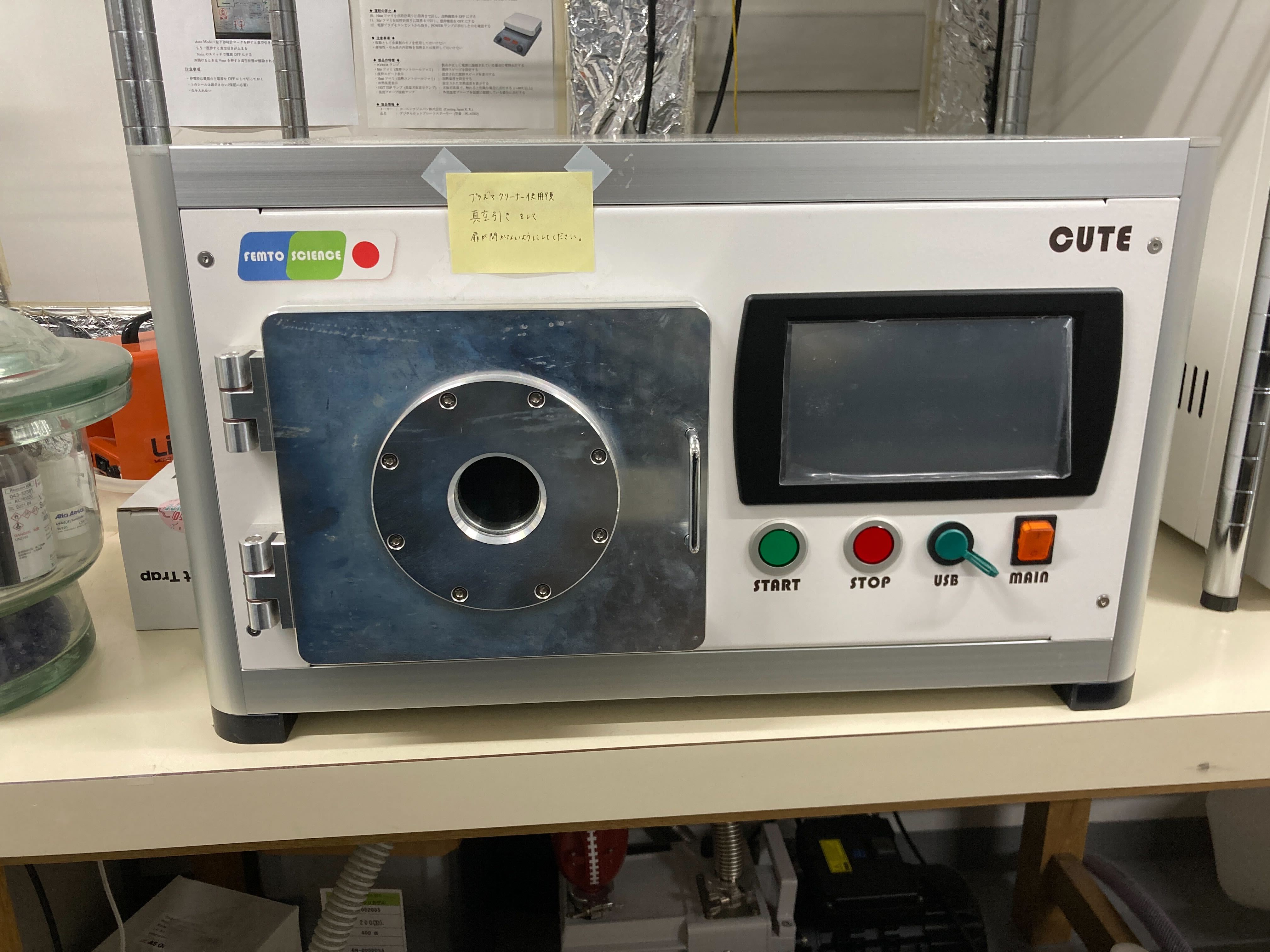
This equipment (Femto Science, CUTE 1MPR) is used to remove organic contaminants and perform surface modification on the substrate for the fabrication of 2D perovskite thin films, for example.