
Principal Spectroscopy Equipment
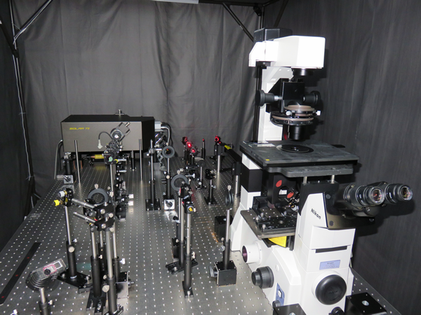 |
Confocal Raman Microspectrometer (632.8 nm excitation) This laboratory-built instrument can measure Raman spectra of the sample such as a living cell under an inverted microscope (Nikon TE2000) with a submicrometer resolution. It uses the 632.8 nm output of a He–Ne laser as the Raman excitation light. The detector used is a TE-cooled CCD detector (Andor iDus DU401-BV) or an electron-multiplying CCD detector (Andor Newton DU970P-BVF). By incorporating volume Bragg grating notch filters that have a very narrow bandwidth, we are able to observe an extremely low-frequency region (down to ±5 cm-1) of Raman spectra. A 3-axis piezoelectric stage (Mad City Labs, Nano-LP100) can be mounted on the microscope stage for Raman imaging experiments. |
|---|---|
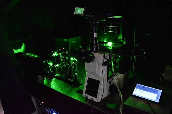 |
Confocal Raman Microspectrometer (532 nm excitation) This Raman microscope is also laboratory-built. It is based on an Olympus inverted microscope (IX73). The excitation laser is an all-solid, optically pumped semiconductor laser (Coherent, Genesis CX532-2000SLM-CDRH), and the detector is an electrically cooled CCD detector (Andor, iDus DU401-BV). Like the 632.8 nm excited Raman microscope, this apparatus is also capable of measuring Raman spectra as low as ±5 cm-1. Using a microscope cooling/heating stage (Linkam, 10021), we can measure Raman spectra at different temperatures (-20 to 120 ℃). |
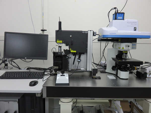 | Nano Raman Imaging Apparatus This state-of-the-art apparatus that combines an atomic force microscope (AFM) and an upright Raman microscope allows not only for Raman measurements synchronized with AFM topographic imaging but also for nanoscale chemical analysis by tip-enhanced Raman scattering (TERS) measurements. 532 nm and 638 nm lasers are implemented for Raman excitation. We use this apparatus to study membrane vesicles produced by bacterial cells as well as thin films of organic–inorganic hybrid perovskites and amyloid fibrils. |
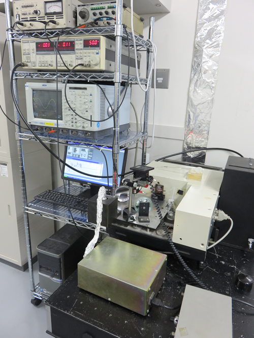 | Infrared Electroabsorption Spectrometer This spectrometer capable of detecting IR absorbance changes of ΔA ≈ 10-8 induced by application of an external electric field is unique to our laboratory. The high sensitivity is achieved by using a dispersive monochromator in combination with AC-coupled amplification, rather than a Fourier transform spectrometer. The detector used is a HgCdTe or an InSb infrared detector. With this apparatus, we are currently investigating the electric field effects of water in reverse micelles and of CH3NH3PbI3 thin films. |
Other Equipment
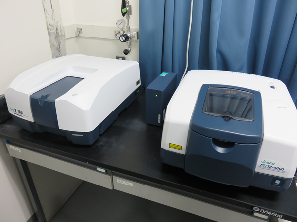 | FTIR Spectrometer (right), UV/VIS Spectrometer (left) These are routinely used for steady-state measurements. An option of attenuated total reflection (ATR) with a ZnSe prism is available for the FTIR spectrometer. |
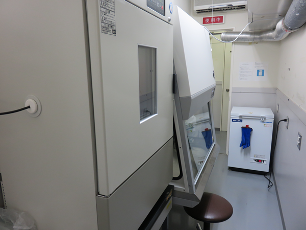 | Cell Culture Facilities Our laboratory has an incubator, a laminar flow cabinet, an autoclave, a deep freezer (-80 °C), and other equipment to culture cells by ourselves (Schizosaccharomyces pombe provided by Prof. Kawamukai of Shimane University; Lactobacillus plantarum, Paracoccus denitrificans, Escherichia coli, etc. provided by Prof. Nomura of University of Tsukuba). |
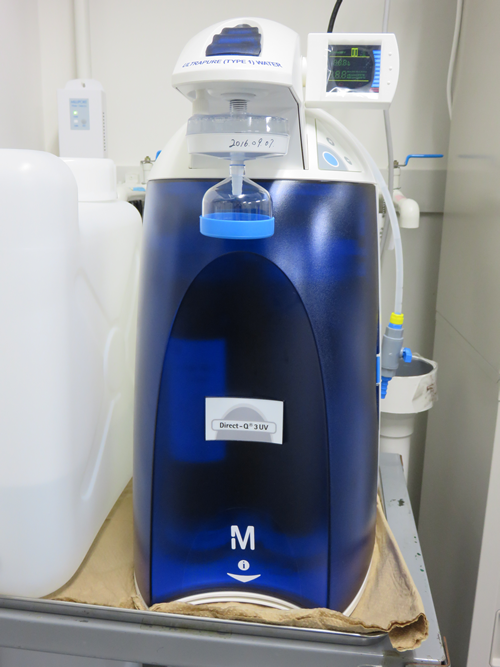 | Ultrapure Water System This apparatus (Merck Millipore Direct-Q UV3) produces ultrapure water from tap water. |
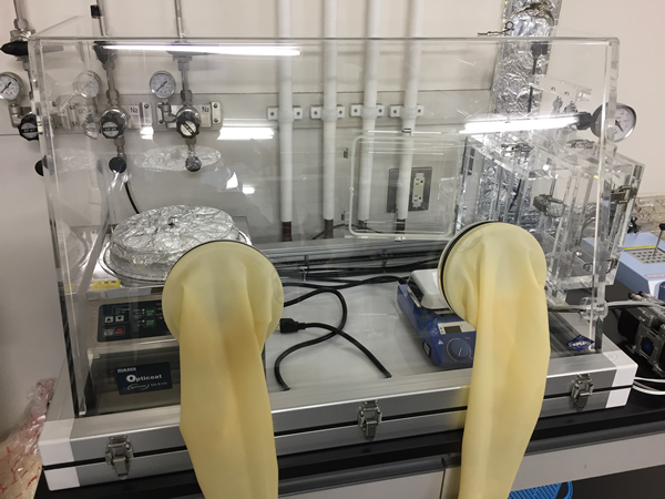 | Glove Box, Spin Coater A nitrogen-filled glove box and a spin coater are used to fabricate organolead halide perovskite thin films. |
|---|