Research Overview
Understanding Materials by Seeing What Happens Inside
Materials and devices surrounding us function through chemical reactions, mass transport, and structural transformations occurring within them. However, these processes rarely proceed uniformly. In many cases, reactions occur locally, evolve dynamically, and exhibit significant spatial and temporal heterogeneity. As a result, conventional analytical techniques that provide only averaged information are often insufficient to reveal the true origins of functionality, degradation, and failure.
At the Matsui Laboratory, we develop and apply advanced X-ray spectroscopic and imaging techniques using high-flux synchrotron radiation at SPring-8, one of the world’s leading synchrotron facilities. Our goal is to visualize chemical-state changes, mass transport, and reaction processes occurring inside materials in a non-destructive, operando, time-resolved, and spatially resolved manner.
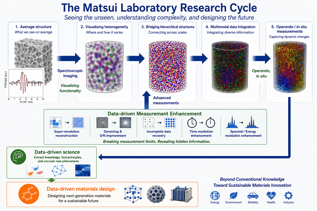
Measurement Technology Development
Many important material functions emerge under non-equilibrium conditions, including gas adsorption, electrochemical reactions, catalytic reactions, adhesion, and fracture processes. Furthermore, these phenomena are often irreversible and may occur only once during the lifetime of a material. For this reason, simply comparing materials before and after operation is often insufficient for understanding the mechanisms responsible for functionality or degradation.
Our laboratory develops operando and in situ measurement techniques that allow us to directly observe chemical-state changes and mass transport while materials are functioning. To achieve this, we design specialized experimental cells and measurement systems that reproduce realistic operating environments while remaining compatible with synchrotron-based X-ray measurements. By bridging laboratory-scale experiments and synchrotron facilities, we aim to observe materials under conditions as close as possible to their actual operating environments.
X-ray Spectroimaging Techniques
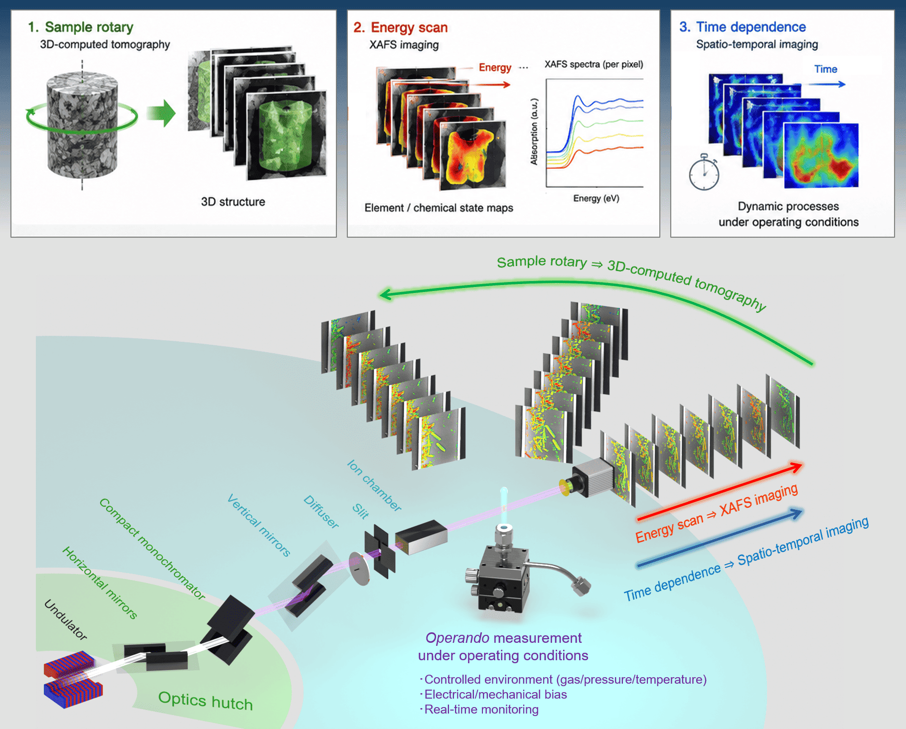
Material degradation and functionality often exhibit strong spatial heterogeneity. However, conventional X-ray measurements generally provide information integrated along the X-ray beam path, making it difficult to determine where reactions occur inside a material.
XAFS-CT imaging combines X-ray Absorption Fine Structure (XAFS) spectroscopy with X-ray computed tomography (CT) to visualize elemental distributions and chemical-state distributions in three dimensions. Our research group has played a leading role in developing this methodology. By combining XAFS spectral analysis with tomographic reconstruction techniques, we can visualize the three-dimensional distributions of chemical states and elements inside complex materials under operating conditions. This technique enables direct observation of hidden reaction pathways and degradation processes occurring within functional materials.
Many important elements in functional materials are present only in trace amounts, making conventional transmission measurements difficult. To address this challenge, we develop high-sensitivity scanning fluorescence XAFS imaging techniques. In this approach, highly focused X-ray beams generated using focusing mirror optics are scanned across the sample surface. Fluorescence X-rays emitted from each position are detected and converted into spatially resolved chemical-state maps.
By repeating measurements across an X-ray absorption edge, two-dimensional XAFS spectroimaging datasets can be acquired and transformed into elemental and chemical-state distributions. Although data acquisition may require several hours or even days, the technique provides exceptional sensitivity and spatial resolution for investigating trace-element behavior.
Many material functions arise from dynamic processes such as adsorption, diffusion, chemical reactions, and degradation. To understand these phenomena, spatial information alone is insufficient. Time evolution must also be captured.
Our laboratory develops time-resolved spectroimaging techniques by selecting characteristic X-ray energies that are particularly sensitive to specific chemical-state changes identified from XAFS spectra. Continuous imaging at these selected energies enables visualization of dynamic chemical processes with high temporal resolution. This approach allows us to simultaneously track the spatial distribution and temporal evolution of chemical-state changes, providing unique insights into reaction mechanisms occurring inside functional materials.
In collaboration with RIKEN and Osaka University, we are developing high-magnification imaging XAFS techniques using Advanced Kirkpatrick–Baez (AKB) mirrors at SPring-8. Unlike conventional diffractive X-ray optics, AKB mirrors utilize total external reflection. Consequently, image position and magnification remain unchanged during energy scans across a wide range of X-ray energies used for XAFS measurements. This unique property eliminates the need for continuous refocusing during XAFS spectroimaging and enables rapid, high-precision spectroscopic imaging with a constant field of view and magnification.
Furthermore, by combining projection X-ray imaging for large-scale observations with AKB-based high-resolution imaging, we are developing multiscale XAFS imaging systems that bridge spatial scales from millimeters to nanometers. This capability allows us to connect local chemical-state changes with the overall behavior of materials, providing a comprehensive understanding of heterogeneous reactions and functional processes.
Functional Materials and Applications
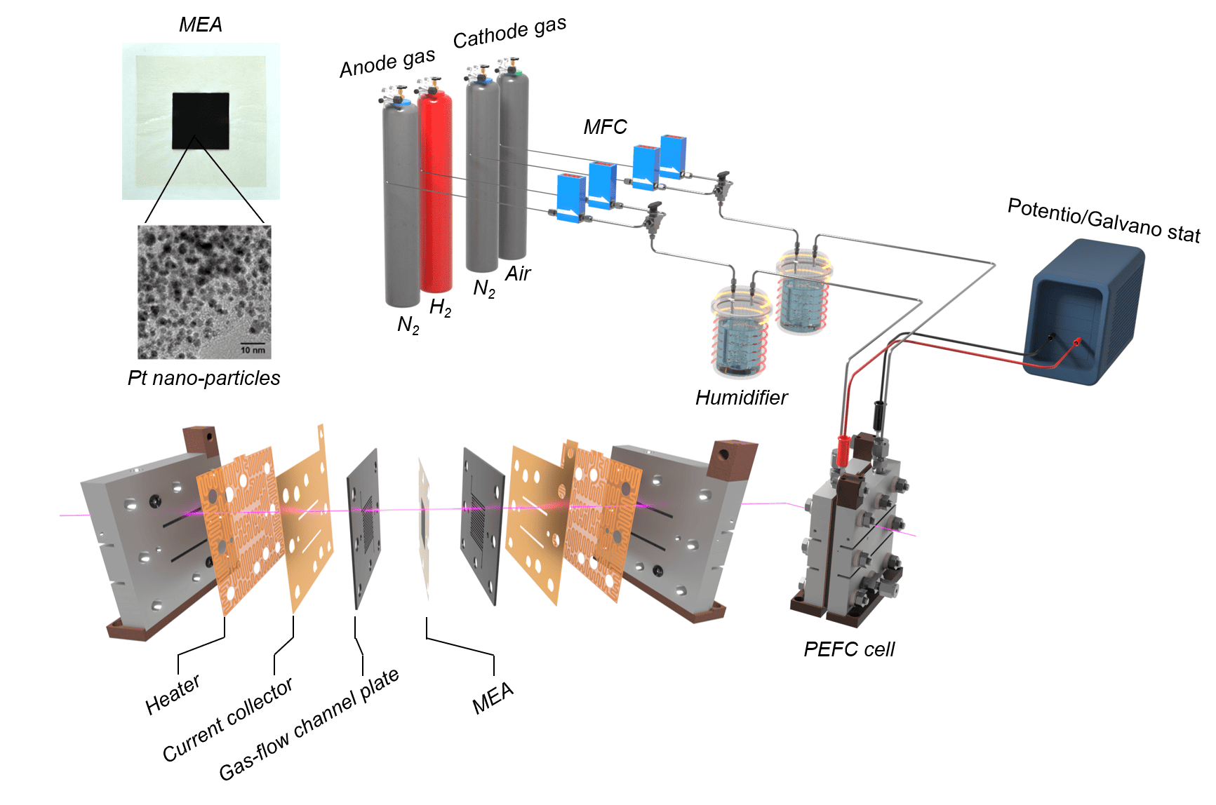
Polymer electrolyte fuel cells (PEFCs) are electrochemical devices that generate electricity from hydrogen and oxygen. As a key technology for realizing a hydrogen-based energy society, PEFCs have already been implemented in commercial fuel-cell vehicles, heavy-duty vehicles, and stationary power systems. Despite these advances, significant challenges remain in improving durability, reducing costs, and expanding applications. Long-term operation often leads to catalyst degradation, poisoning by impurities, and complex conditioning phenomena that can significantly affect performance.
Our laboratory utilizes advanced XAFS imaging techniques to visualize chemical-state changes within operating PEFCs under realistic conditions. By reproducing degradation, poisoning, and conditioning processes in operando settings, we investigate the origins of performance loss and identify the mechanisms underlying functional deterioration.
Sel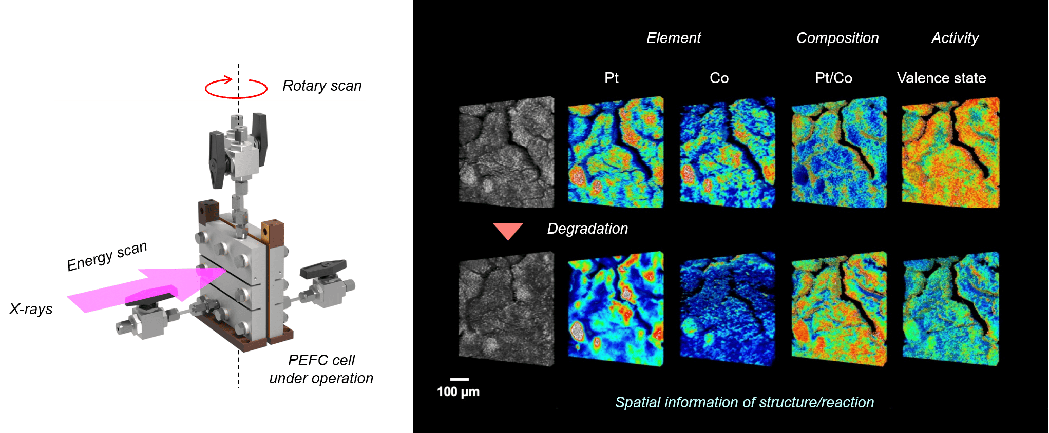 ected Publications
- Phys. Chem. Chem. Phys. 2024, 26, 15115-15119. https://pubs.rsc.org/en/content/articlelanding/2024/cp/d4cp00728j
- Catal. Sci. Technol. 2024, 14, 1501-1511. https://pubs.rsc.org/en/content/articlelanding/2024/cy/d3cy01438j#!divCitation
- Catal. Sci. Technol. 2023, 13, 4360-4366. https://pubs.rsc.org/en/content/articlelanding/2023/cy/d3cy00241a
- ACS Appl. Mater. Interfaces 2022, 14, 6762-6776. https://pubs.acs.org/doi/10.1021/acsami.1c22336
- ChemNanoMat. 2022, 8, e202200008. https://aces.onlinelibrary.wiley.com/doi/10.1002/cnma.202200008
- J. Phys. Chem. C 2019, 123, 18844-18853. https://pubs.acs.org/doi/10.1021/acs.jpcc.9b05005
- Chem. Rec. 2019, 19, 1380-1392. https://onlinelibrary.wiley.com/doi/abs/10.1002/tcr.201800123
- J. Phys. Chem. 2018, 122, 14511-14517. https://pubs.acs.org/doi/10.1021/acs.jpcc.8b02541
- Angew. Chem. Int. Ed. 2017, 56, 9371-9375. https://onlinelibrary.wiley.com/doi/10.1002/anie.201703940
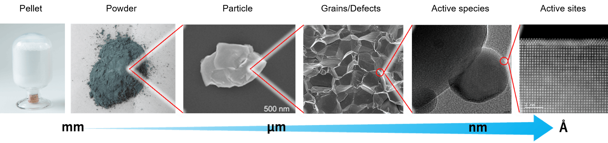
Solid catalysts possess highly hierarchical structures spanning multiple length scales, from centimeter and millimeter-scale pellets and powder to atomic-scale active sites. Although catalytic performance ultimately originates from local chemical reactions occurring at active sites, the relationship between local phenomena and macroscopic catalyst performance remains poorly understood.
Furthermore, catalysts often operate under highly dynamic and non-equilibrium conditions. Active metal species may undergo oxidation, reduction, migration, aggregation, or restructuring during operation. Simultaneously, reactants and products continuously diffuse through porous structures, creating complex reaction environments that evolve in both space and time.
Our laboratory develops and applies advanced XAFS imaging techniques to visualize these dynamic processes directly. By observing chemical-state distributions, reaction propagation, and mass-transport phenomena within catalyst materials, we seek to identify the true factors controlling catalytic activity, selectivity, stability, and deactivation. Current research topics include fuel-cell catalysts, automotive exhaust-gas purification catalysts, and metal-supported catalysts.
Selected Public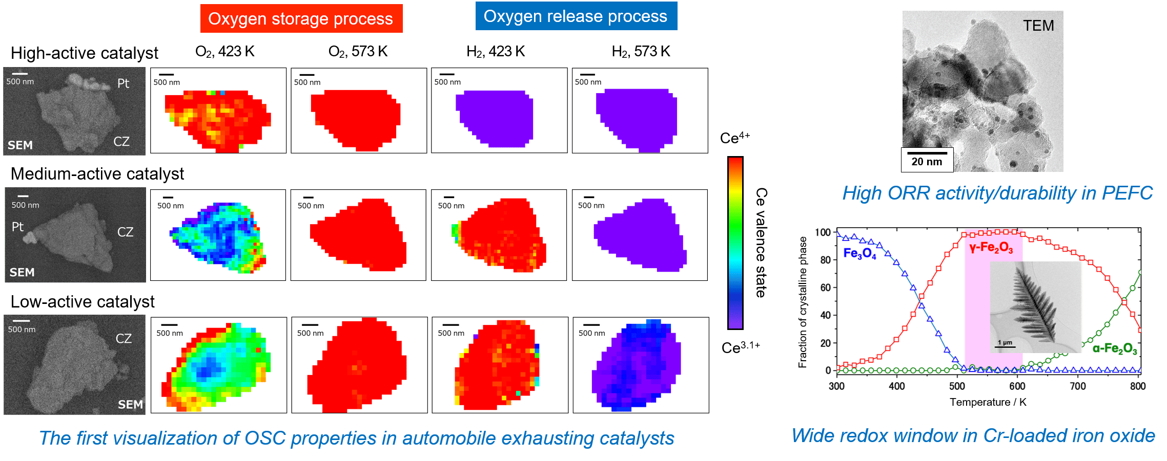 ations
- Phys. Chem. Chem. Phys. 2023, 25, 17197. https://pubs.rsc.org/en/content/articlelanding/2023/cp/d3cp00907f
- Phys. Chem. Chem. Phys. 2020, 22, 28093-28099. https://pubs.rsc.org/en/content/articlelanding/2020/cp/d0cp04416d
- Angew. Chem. Int. Ed. 2016, 55, 12022-12025. https://onlinelibrary.wiley.com/doi/abs/10.1002/anie.201606046
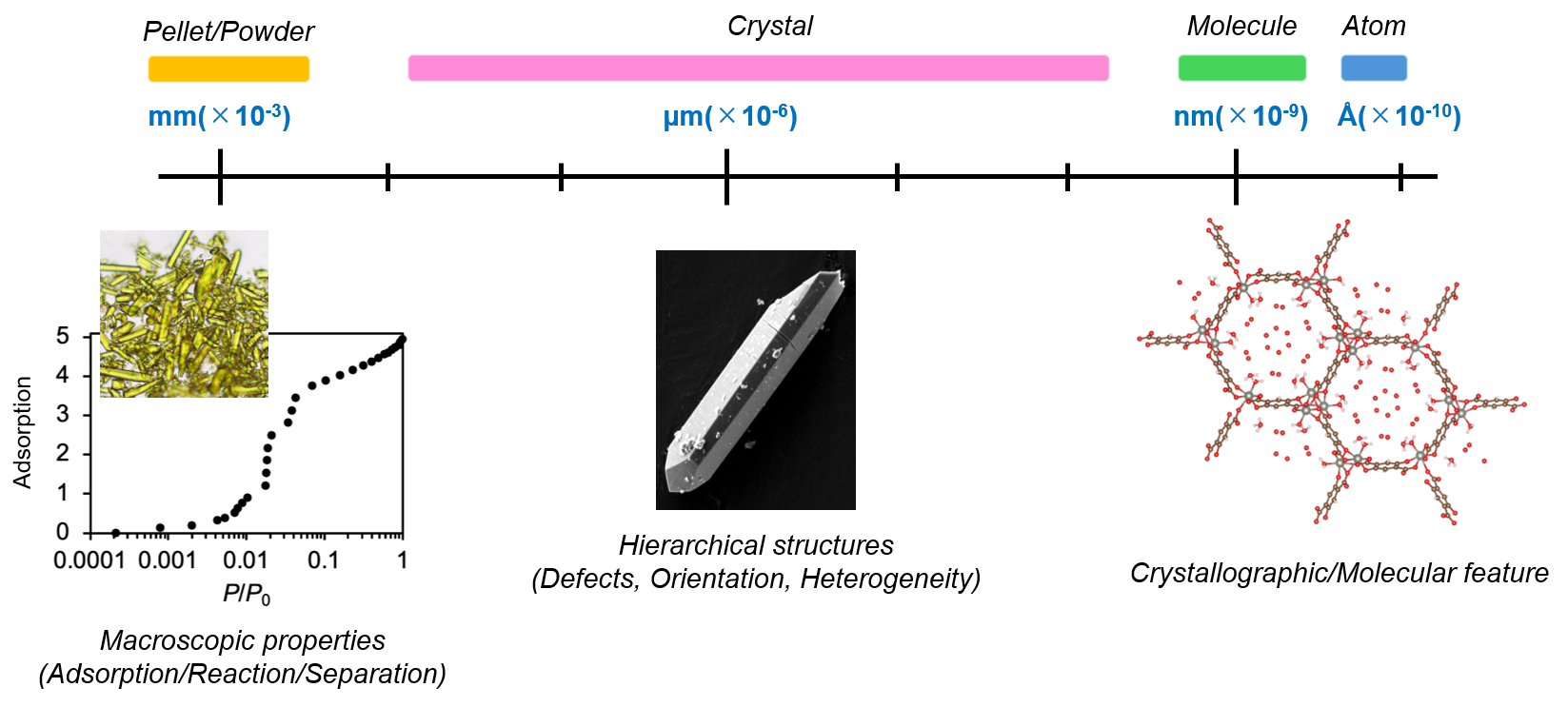
Metal–Organic Frameworks (MOFs) are porous crystalline materials constructed from metal ions and organic ligands. Their exceptionally high surface areas and tunable pore structures have attracted tremendous attention for applications such as gas storage, gas separation, sensing, and energy technologies.
The scientific understanding of MOFs has traditionally been driven by crystallographic studies that reveal molecular-level structures. However, many important questions remain unanswered. How does adsorption propagate through a crystal? How do guest molecules diffuse through complex pore networks? How do structural transformations evolve during adsorption? Why do nominally identical crystals sometimes exhibit different adsorption behaviors?
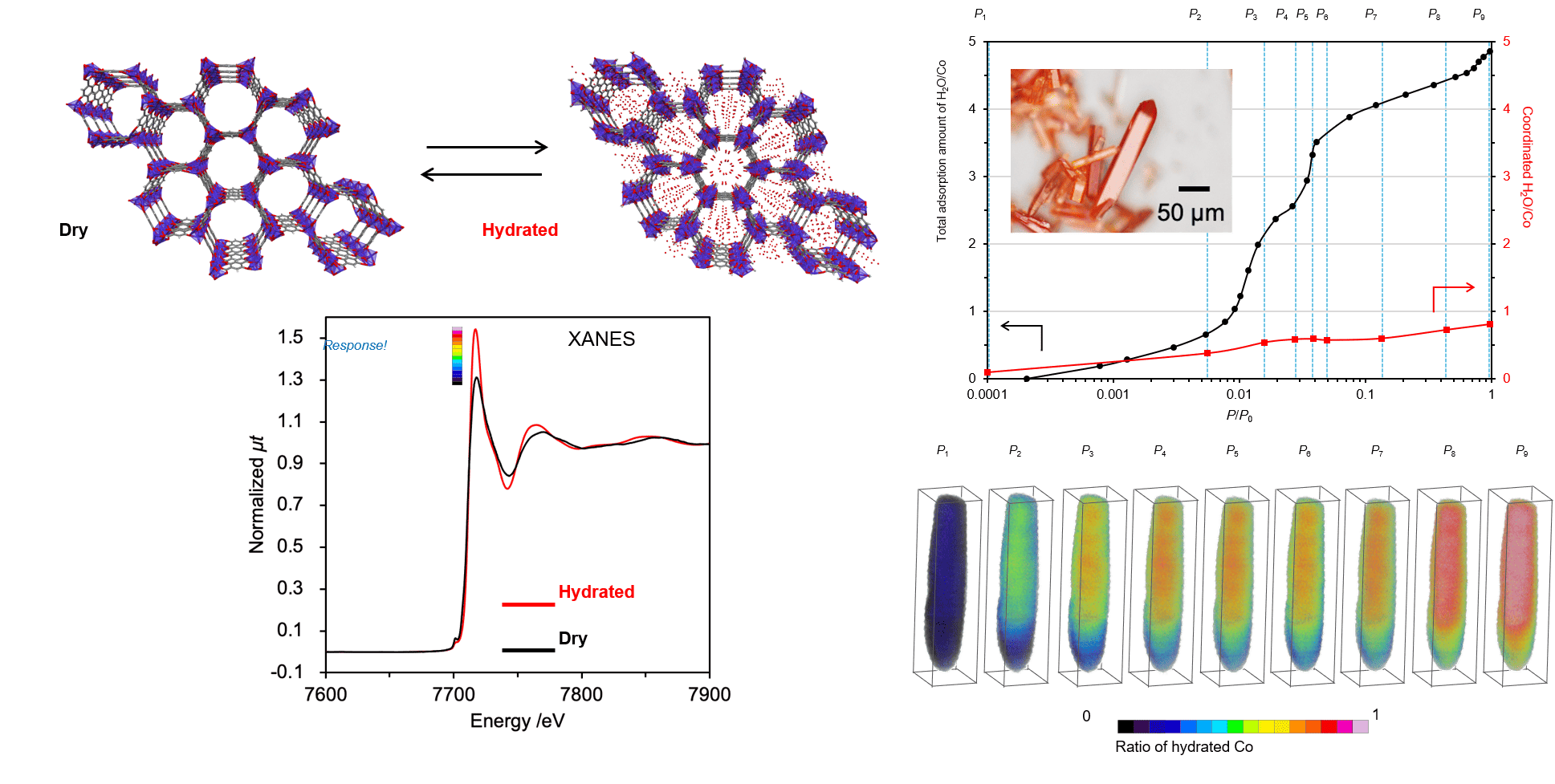
To address these challenges, our laboratory applies advanced X-ray spectroimaging techniques to directly visualize adsorption and diffusion processes within individual MOF crystals. By tracking the spatial and temporal evolution of local chemical states and mass transport during gas adsorption, we investigate the influence of crystal morphology, defects, diffusion pathways, and host–guest interactions on functional performance.
 Selected Publications
- J. Am. Chem. Soc. 2024, 146, 13, 9181-9190. https://pubs.acs.org/doi/10.1021/jacs.3c14778

The performance and reliability of many industrial products depend critically on the integrity of buried interfaces between dissimilar materials. In automobile tires, for example, strong adhesion between rubber and steel cords is essential for ensuring safety, durability, and long-term performance.
Despite their technological importance, buried interfaces remain among the most difficult regions of materials to investigate. Conventional studies typically evaluate adhesion through mechanical testing and post-mortem analysis after failure has occurred. While such approaches provide valuable information regarding the final state of a material, they often fail to reveal how interfacial reactions evolve during operation, how degradation initiates, and why failure ultimately occurs.
Our laboratory seeks to bridge this gap by combining advanced X-ray spectroscopic imaging techniques with mechanical testing. We directly visualize chemical reactions occurring at buried rubber–metal interfaces while simultaneously monitoring the mechanical response of the interface. Particular attention is devoted to sulfurization reactions, chemical-state evolution of metallic components, moisture-induced degradation, and other interfacial processes that govern adhesion performance between rubber and metal. By observing these reactions, we investigate how local chemical transformations influence interfacial strength and durability.
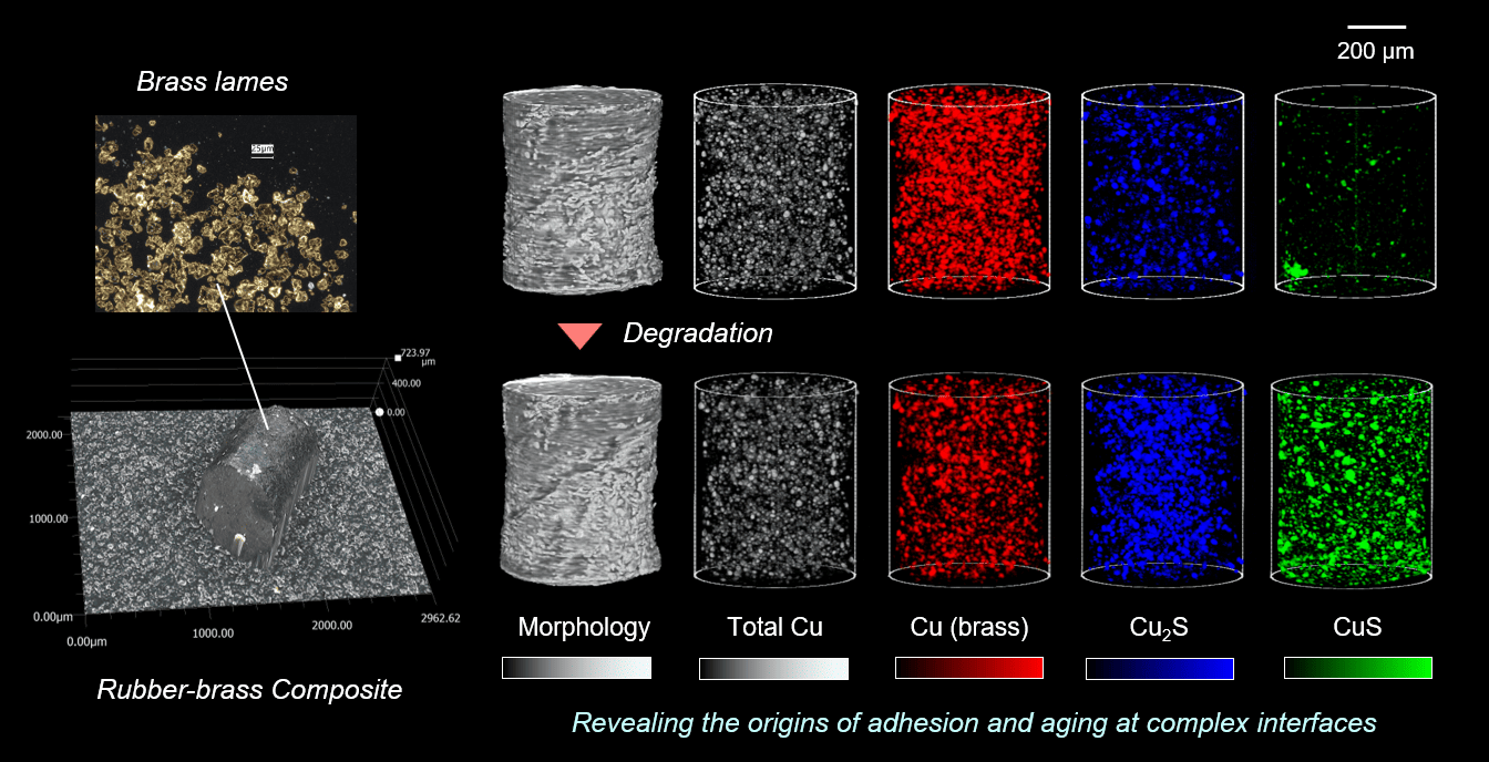
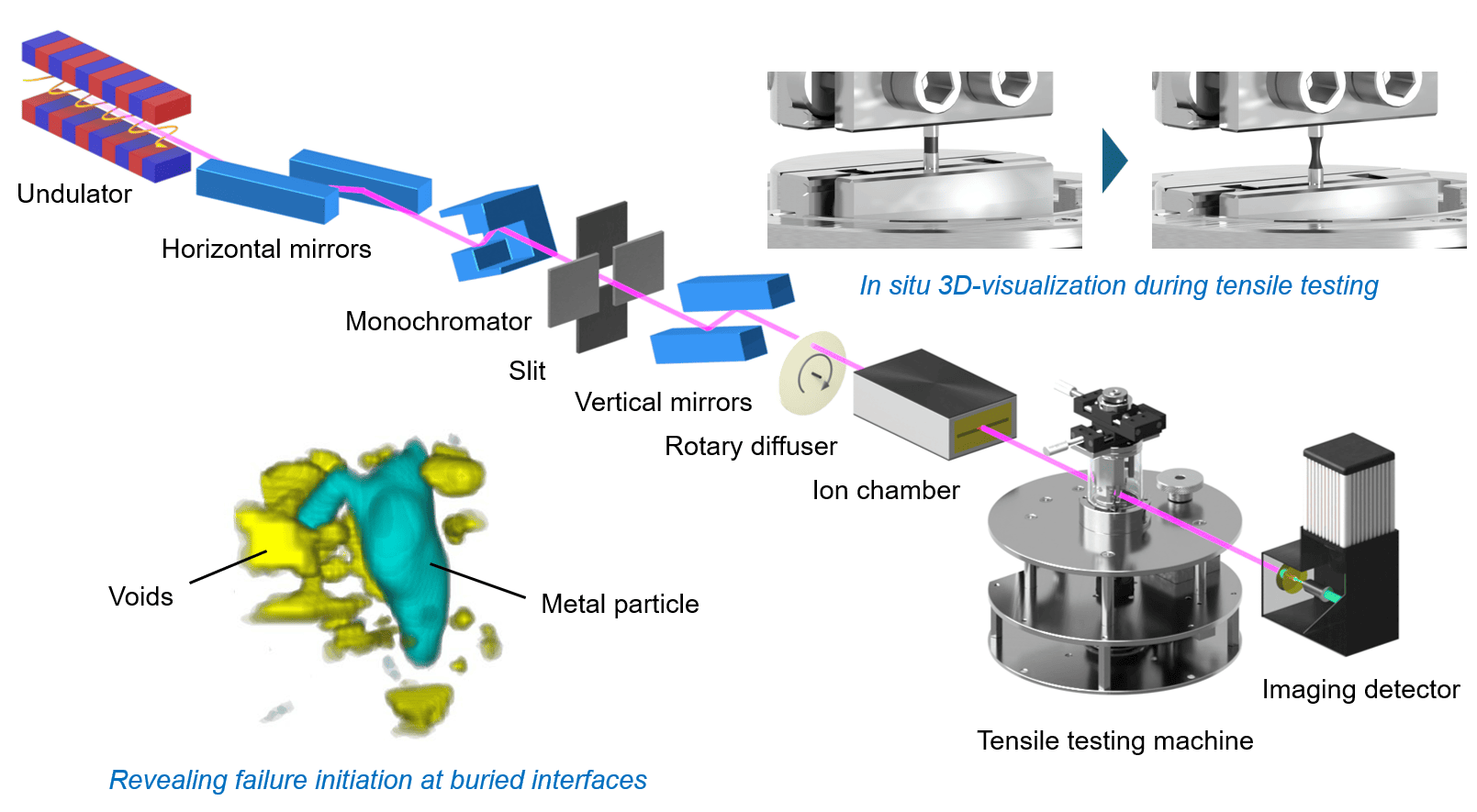
Furthermore, by integrating tensile and delamination testing with our synchrotron-based imaging techniques, we visualize the initiation and propagation of damage during mechanical loading. This approach enables us to identify where failure originates, how cracks develop, and how interfacial chemistry influences interfacial fracture behavior across multiple length scales. Through these studies, we aim to establish a direct relationship between chemical reactions, interfacial structure, mechanical properties, and failure processes. Ultimately, our goal is to develop a fundamental scientific framework that connects chemistry and mechanics, providing new design principles for durable adhesion technologies and multifunctional materials.
 Selected Publications
- Communications Materials 2023, 4, 88. https://www.nature.com/articles/s43246-023-00413-z